The ‘myth’ of the pterosaur uropatagium: now a hoax

Hone and Prondvai 2025
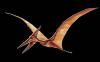
focus this studiy on the purported, but still invalid, uropatagium impossibly stretched between the legs and lateral toe tips of long-tailed basal pterosaurs like Sordes (Figs 1, 3, 4). No outgroup taxa share this trait. Rather, bipedal Sharovipteryx (Fig 5) and Cosesaurus (Fig 2), preserve a uropatagial membrane behind each leg – as also preserved in later pterosaurs, like Jeholopterus (Fig 7) and Pterodactylus (Fig 8). The authors wrote, “pterodactyloid pterosaurs had a greatly reduced and split uropatagium spanning a much smaller triangular area between the knee joint and the base of the tail.”
Not split. The post hind limb membrane was always the same from pre-pterosaurs (Figs 2, 5) to the later pterosaurs (Peters 2002, Figs 7, 8), always behind each hind limb – never stretched from lateral toe tip to lateral toe tip.
That would have been such a bizarre structure – which is why the central spanning uropatagium hypothesis has now moved from myth to hoax. This is not the first of Dr David Hone’s blunders. It follows a long list linked here.
Figure 1. Sordes interpreted by me in Nature 1995. At that time. remember, no one had ever heard of a uropatagium. And the purported “fact” that this flap of skin spanned the hind limbs without connecting to the tail seemed pretty hard to swallow for a majority of paleontologists. The only images available were small and indistinct. Even so, an attempt was made here to understand the taphonomy of the specimen and how it came to sport such a strange autapomorphy that has not been seen since on any pterosaur fossil. Despite the sincerity of this effort, it includes several mistakes rectified now in ReptileEvolution.com/sordes.htm after publication of the specimen in a larger format with higher resolution.
” data-medium-file=”https://pterosaurheresies.wordpress.com/wp-content/uploads/2012/07/petes-1995-reinterpretation-wing-shape-in-sordes-july-2012-tiny.jpg?w=300″ data-large-file=”https://pterosaurheresies.wordpress.com/wp-content/uploads/2012/07/petes-1995-reinterpretation-wing-shape-in-sordes-july-2012-tiny.jpg?w=584″ class=”size-full wp-image-6510″ src=”https://pterosaurheresies.wordpress.com/wp-content/uploads/2012/07/petes-1995-reinterpretation-wing-shape-in-sordes-july-2012-tiny.jpg” alt=”" width=”584″ height=”373″ srcset=”https://pterosaurheresies.wordpress.com/wp-content/uploads/2012/07/petes-1995-reinterpretation-wing-shape-in-sordes-july-2012-tiny.jpg?w=584&h=373 584w, https://pterosaurheresies.wordpress.com/wp-content/uploads/2012/07/petes-1995-reinterpretation-wing-shape-in-sordes-july-2012-tiny.jpg?w=150&h=96 150w, https://pterosaurheresies.wordpress.com/wp-content/uploads/2012/07/petes-1995-reinterpretation-wing-shape-in-sordes-july-2012-tiny.jpg?w=300&h=192 300w, https://pterosaurheresies.wordpress.com/wp-content/uploads/2012/07/petes-1995-reinterpretation-wing-shape-in-sordes-july-2012-tiny.jpg 600w” sizes=”(max-width: 584px) 100vw, 584px” />
Figure 1. Sordes interpreted by David Peters in Nature 1995. At that time. remember, no one had ever heard of a uropatagium. And the purported “fact” that this flap of skin spanned the hind limbs without connecting to the tail seemed pretty hard to swallow for a majority of paleontologists. The only images available were small and indistinct. Even so, an attempt was made here to understand the taphonomy of the specimen and how it came to sport such a strange autapomorphy that has not been seen since on any pterosaur fossil. Despite the sincerity of this effort, it includes several mistakes rectified now in ReptileEvolution.com/sordes.htm after publication of the specimen in a larger format with higher resolution.
The authors wrote,
“Although pterosaurs have undergone something of a recent renaissance in research, many basic features of their biology including their origins, functional morphology and palaeoecology are still strongly debated.”
Not ‘debated’. Omitted. Ignored. If debated Hone and Prondvai should have explored all omitted citations then presented evidence supporting their side while falsifying opposing evidence. As you’ll see, all three of their best examples either fail to understand taphonomic displacement or fail to present evidence of a uropatagium spanning the lateral pedal digits and hind limbs.
You’ll find workers, like Hone and Prondvai, using terms like
‘debated’ and ‘disagreement’ as if data were based on opinions, instead of something substantial like: ‘author X reported data. Here we falsify that hypothesis/observation with more precise data.’ Unfortunately that method doesn’t happen here. Instead Hone and Prodvai use cartoon = freehand pterosaur diagrams and ignore opposing citations that employed and interpreted data more precisely.
Figure 6. The uropatagium following the left hind limb of Cosesaurus, photographed by Ellenberger (1993).
” data-medium-file=”https://pterosaurheresies.wordpress.com/wp-content/uploads/2012/03/ellencosesaurus-urop.jpg?w=300″ data-large-file=”https://pterosaurheresies.wordpress.com/wp-content/uploads/2012/03/ellencosesaurus-urop.jpg?w=584″ class=”size-full wp-image-5289″ src=”https://pterosaurheresies.wordpress.com/wp-content/uploads/2012/03/ellencosesaurus-urop.jpg” alt=”The uropatagium following the left hind limb of Cosesaurus” width=”584″ height=”262″ srcset=”https://pterosaurheresies.wordpress.com/wp-content/uploads/2012/03/ellencosesaurus-urop.jpg?w=584&h=262 584w, https://pterosaurheresies.wordpress.com/wp-content/uploads/2012/03/ellencosesaurus-urop.jpg?w=150&h=67 150w, https://pterosaurheresies.wordpress.com/wp-content/uploads/2012/03/ellencosesaurus-urop.jpg?w=300&h=135 300w, https://pterosaurheresies.wordpress.com/wp-content/uploads/2012/03/ellencosesaurus-urop.jpg 588w” sizes=”(max-width: 584px) 100vw, 584px” />
Figure 2. The uropatagium following the left hind limb of Cosesaurus, photographed by Ellenberger (1993). This middle Triassic taxon was ancestral to Sharovipteryx and pterosaurs in the LRT.
The authors wrote,
“This review presents the current knowledge of the shape, extent, structure and function of the uropatagium that lies between the legs in pterosaurs, based on the available fossil evidence. Both direct evidence from soft tissue preservation and indirect evidence from osteological and ichnological data, suggest variation in the uropatagium among taxa, although evolutionary trends can still be outlined.”
Ichnological data does not and cannot portray whether or not a trackmaker had uropatagia, frills, a gular sac and other extradermal membranes.
Use of the word ‘suggest’ means whatever follows is a biased opinion.
Better to rely on hard evidence when dealing with soft tissue.
The Sordes uropatagium is actually displaced wing material carried between the ankles by the displaced radius and ulna.
” data-medium-file=”https://pterosaurheresies.wordpress.com/wp-content/uploads/2011/07/sordes-uropatagia.jpg?w=300″ data-large-file=”https://pterosaurheresies.wordpress.com/wp-content/uploads/2011/07/sordes-uropatagia.jpg?w=584″ class=”size-full wp-image-149″ src=”https://pterosaurheresies.wordpress.com/wp-content/uploads/2011/07/sordes-uropatagia.jpg” alt=”The myth of the pterosaur uropatagium” width=”584″ height=”459″ srcset=”https://pterosaurheresies.wordpress.com/wp-content/uploads/2011/07/sordes-uropatagia.jpg?w=584&h=459 584w, https://pterosaurheresies.wordpress.com/wp-content/uploads/2011/07/sordes-uropatagia.jpg?w=150&h=118 150w, https://pterosaurheresies.wordpress.com/wp-content/uploads/2011/07/sordes-uropatagia.jpg?w=300&h=236 300w, https://pterosaurheresies.wordpress.com/wp-content/uploads/2011/07/sordes-uropatagia.jpg?w=768&h=603 768w, https://pterosaurheresies.wordpress.com/wp-content/uploads/2011/07/sordes-uropatagia.jpg 900w” sizes=”(max-width: 584px) 100vw, 584px” />
Figure 3. The Sordes uropatagium is actually displaced wing material carried between the ankles by the displaced radius and ulna.
Unwin 1999 invented the term
‘cruropatagium‘ for an interpretation of a membrane stretching between the crus = tibia + fibula of the hind limbs (plus the lateral toe tips) in Sordes (Fig 4) first described by Sharov 1971, who also misinterpreted the specimen. He was an insect specialist.
When attendees were presented the purported uropatagium in Sordes
described by David Unwin at a SVP conference, friends looked at each other and wondered, “How is that possible?” Not only does such a uropatagium inhibit and affect defecation and reproduction, but terrestrial locomotion – and none of us could figure out how such a structure spanning opposite toe tips would or could develop either phylogenetically or ontogenetically.
It made no sense then – or now.
Figure 1. Uropatagium of Sordes according to Sharov 1971 and Unwin/Bakhurina 1994.
” data-medium-file=”https://pterosaurheresies.wordpress.com/wp-content/uploads/2011/07/sordessharov2.jpg?w=134″ data-large-file=”https://pterosaurheresies.wordpress.com/wp-content/uploads/2011/07/sordessharov2.jpg?w=209″ class=”size-full wp-image-153″ src=”https://pterosaurheresies.wordpress.com/wp-content/uploads/2011/07/sordessharov2.jpg” alt=”Uropatagium of Sordes according to Sharov 1971 and Unwin/Bakhurina 1994.” width=”209″ height=”469″ srcset=”https://pterosaurheresies.wordpress.com/wp-content/uploads/2011/07/sordessharov2.jpg 209w, https://pterosaurheresies.wordpress.com/wp-content/uploads/2011/07/sordessharov2.jpg?w=67&h=150 67w” sizes=”(max-width: 209px) 100vw, 209px” />
Figure 4. Uropatagium of Sordes according to Sharov 1971 and Unwin/Bakhurina 1994.
Hone and Prondvai wrote,
“We prefer to use the term uropatagium in pterosaurs and define it as a flight membrane attaching to the hind limbs and to the fifth toe of the pes (in forms where it is present) and, additionally, most probably the tail (or at least the base of the tail).”
So they are doubling down on a myth.
Their paper presents a short history of the uropatagium, abridged here:
Hone and Prondvai wrote,
“Owen (1870) proposed that the fifth toe in pterosaurs might have had the same function as the calcar in bats, namely, to provide control and structural support for the uropatagium. One of the consequences of this, again based on observations of extant bats, is that the integration of the hind limb within the flight membranes might have made terrestrial locomotion more complicated for pterosaurs (Abel 1925).”
Owen 1870 had only pterosaurs at hand with this odd fifth toe. He could not compare the identical structure to Cosesaurus (Fig 2, the maker of Rotodactylus ichnites), Sharovipteryx (Fig 5), Langobardisaurus, Tanytrachelos and the largest tanystropheids. These taxa are omitted from Hone and Prondvai 2025. Their omission is a pattern among pterosaur workers.
Figure 3. Sharovipteryx reconstructed. Note the flattened torso.
” data-medium-file=”https://pterosaurheresies.wordpress.com/wp-content/uploads/2018/02/sharovipteryx-recon588.jpg?w=250″ data-large-file=”https://pterosaurheresies.wordpress.com/wp-content/uploads/2018/02/sharovipteryx-recon588.jpg?w=584″ class=”size-full wp-image-29678″ src=”https://pterosaurheresies.wordpress.com/wp-content/uploads/2018/02/sharovipteryx-recon588.jpg” alt=”Figure 3. Sharovipteryx reconstructed. Note the flattened torso.” width=”584″ height=”701″ srcset=”https://pterosaurheresies.wordpress.com/wp-content/uploads/2018/02/sharovipteryx-recon588.jpg?w=584&h=701 584w, https://pterosaurheresies.wordpress.com/wp-content/uploads/2018/02/sharovipteryx-recon588.jpg?w=125&h=150 125w, https://pterosaurheresies.wordpress.com/wp-content/uploads/2018/02/sharovipteryx-recon588.jpg?w=250&h=300 250w, https://pterosaurheresies.wordpress.com/wp-content/uploads/2018/02/sharovipteryx-recon588.jpg 588w” sizes=”(max-width: 584px) 100vw, 584px” />
Figure 5. Sharovipteryx reconstructed. Note the flattened torso. Note the tanystropheid pedal digit 5 supporting each uropatagium extended to all the toes.
Hone and Prondvai wrote,
“Stromer (1913), contested this model saying that the hind limbs were free of the flight membranes and that pterosaurs lacked a uropatagium.”
Stromer was writing about Rhamphorhynchus gemmingi (Fig 6).
Figure 1. Click to enlarge. Specimens nesting with Rhamphorhynchus gemmingi in phylogenetic order from left to right, n43, n74 (the holotype), n38, n75 and n52, otherwise known as R. megadactylus.
” data-medium-file=”https://pterosaurheresies.wordpress.com/wp-content/uploads/2012/05/rhamph-gemmingi-clade-588.jpg?w=300″ data-large-file=”https://pterosaurheresies.wordpress.com/wp-content/uploads/2012/05/rhamph-gemmingi-clade-588.jpg?w=584″ class=”size-full wp-image-5925″ src=”https://pterosaurheresies.wordpress.com/wp-content/uploads/2012/05/rhamph-gemmingi-clade-588.jpg” alt=”Specimens nesting with Rhamphorhynchus gemmingi” width=”584″ height=”187″ srcset=”https://pterosaurheresies.wordpress.com/wp-content/uploads/2012/05/rhamph-gemmingi-clade-588.jpg?w=584&h=187 584w, https://pterosaurheresies.wordpress.com/wp-content/uploads/2012/05/rhamph-gemmingi-clade-588.jpg?w=1168&h=374 1168w, https://pterosaurheresies.wordpress.com/wp-content/uploads/2012/05/rhamph-gemmingi-clade-588.jpg?w=150&h=48 150w, https://pterosaurheresies.wordpress.com/wp-content/uploads/2012/05/rhamph-gemmingi-clade-588.jpg?w=300&h=96 300w, https://pterosaurheresies.wordpress.com/wp-content/uploads/2012/05/rhamph-gemmingi-clade-588.jpg?w=768&h=246 768w, https://pterosaurheresies.wordpress.com/wp-content/uploads/2012/05/rhamph-gemmingi-clade-588.jpg?w=1024&h=328 1024w” sizes=”(max-width: 584px) 100vw, 584px” />
Figure 6. Specimens nesting with Rhamphorhynchus gemmingi in phylogenetic order from left to right, n43, n74 (the holotype), n38, n75 and n52, otherwise known as R. megadactylus.
Hone and Prondvai wrote,
“Padian (1979, 1983a, b, 1985, 1991, 2003)… reconstructed pterosaurs as bipedal, digitigrade, cursorial archosaurs with the hind limbs free of both the brachiopatagium and uropatagium. This bipedal reconstruction also challenged the notion that the fifth toe controlled a uropatagium, and rather suggested that the toe was a pivot during walking. In addition, the fact that more derived pterodactyloids have only a reduced fifth toe was used as an argument against the uropatagium.”
In Padian 1983 (Fig 9, re-describing Dimorphodon) pedal digit 5 was not used as a pivot, whatever ‘pivot’ means in this context. Padian did not know what to do with pedal digit 5, so he let it just be there, swinging freely. Later, Peters 2000a matched pedal digit 5 in Cosesaurus to previously unexplained Rotodactylus tracks in which pedal digit 5 made a small circular impression, like a high heel, in a digitigrade and sometimes bipedal set of ichnites. This citation and data were omitted/ignored by Hone and Prondvai.
Other physical evidence of a uropatagium.
“These include the exquisitely preserved holotype specimen of the anurognathid Jeholopterus ningchengensis IVPP V12705 (Wang et al. 2002, Kellner et al. 2009), another anurognathid, Luopterus mutoudengensis (JZMP-04-07-3) with a poorly preserved uropatagium was also subsequently described (Lü & Hone 2012).
Jeholopterus (image from Hone and Prondvai, shown in Fig 7) is an excellent example of a LACK of a uropatagium stretched between the hind limbs and lateral toes. It was featured in Peters 2002, a citation omitted from Hone and Prondvai.
When authors invite you to see something that is clearly not there, in such a boldface manner, that qualifies as a hoax.
Figure x. From Hone and Prondvai showing the hind limbs of Jeholopterus LACKING a central uropatagium between the lateral pedal digits.
” data-medium-file=”https://pterosaurheresies.wordpress.com/wp-content/uploads/2025/09/jeholopterus.hindlimbs588.gif?w=257″ data-large-file=”https://pterosaurheresies.wordpress.com/wp-content/uploads/2025/09/jeholopterus.hindlimbs588.gif?w=584″ class=”size-full wp-image-94562″ src=”https://pterosaurheresies.wordpress.com/wp-content/uploads/2025/09/jeholopterus.hindlimbs588.gif” alt=”Figure x. From Hone and Prondvai showing the hind limbs of Jeholopterus LACKING a central uropatagium between the lateral pedal digits. ” width=”584″ height=”682″ srcset=”https://pterosaurheresies.wordpress.com/wp-content/uploads/2025/09/jeholopterus.hindlimbs588.gif?w=584&h=682 584w, https://pterosaurheresies.wordpress.com/wp-content/uploads/2025/09/jeholopterus.hindlimbs588.gif?w=128&h=150 128w, https://pterosaurheresies.wordpress.com/wp-content/uploads/2025/09/jeholopterus.hindlimbs588.gif?w=257&h=300 257w, https://pterosaurheresies.wordpress.com/wp-content/uploads/2025/09/jeholopterus.hindlimbs588.gif 588w” sizes=”(max-width: 584px) 100vw, 584px” />
Figure 7. From Hone and Prondvai showing the hind limbs of Jeholopterus LACKING a central uropatagium between the lateral pedal digits.
Luopterus
was originally named Dendrorhynchoides (Fig 10). The uropatagium is the standard type behind each leg. So, another hoax.
scolopaciceps BSP 1937 I 18 (Broili 1938, P. kochi No. 21 of Wellnhofer 1970, 1991)
” data-medium-file=”https://pterosaurheresies.wordpress.com/wp-content/uploads/2025/09/pterodactylus.n21-588.gif?w=300″ data-large-file=”https://pterosaurheresies.wordpress.com/wp-content/uploads/2025/09/pterodactylus.n21-588.gif?w=584″ class=”wp-image-94558 size-full” src=”https://pterosaurheresies.wordpress.com/wp-content/uploads/2025/09/pterodactylus.n21-588.gif” alt=”Figure x. From Hone and Prondvai showing the hind limb of Pterodactylus scolopaciceps BSP 1937 I 18 (Broili 1938, P. kochi No. 21 of Wellnhofer 1970, 1991) This shows the typical orientation of each uropatagium in pterosaurs, Cosesaurus and Sharovipteryx: behind each leg, deepest behind the knee. ” width=”584″ height=”556″ srcset=”https://pterosaurheresies.wordpress.com/wp-content/uploads/2025/09/pterodactylus.n21-588.gif?w=584&h=556 584w, https://pterosaurheresies.wordpress.com/wp-content/uploads/2025/09/pterodactylus.n21-588.gif?w=150&h=143 150w, https://pterosaurheresies.wordpress.com/wp-content/uploads/2025/09/pterodactylus.n21-588.gif?w=300&h=286 300w, https://pterosaurheresies.wordpress.com/wp-content/uploads/2025/09/pterodactylus.n21-588.gif 588w” sizes=”(max-width: 584px) 100vw, 584px” />
Figure 8. From Hone and Prondvai showing the hind limb of Pterodactylus scolopaciceps BSP 1937 I 18 (Broili 1938, P. kochi No. 21 of Wellnhofer 1970, 1991) This shows the typical orientation of each uropatagium in pterosaurs, Cosesaurus and Sharovipteryx: behind each leg, deepest behind the knee.
Hone and Prondvai wrote,
“Evidence for a uropatagium is also present in pterodactyloids. The “Munich specimen” (Broili 1938 – BSPG 1937. I. 18) of Pterodactylus shows one.”
Actually (Fig 8, from Hone and Prondvai) the published photo shows a standard uropatagium behind one leg, as in Cosesaurus (Fig 2), Sharovipteryx and all other pterosaurs, including Sordes. The other leg is below the rest of the specimen.
“A second uropatagium morphotype is reported from two specimens of Pterodactylus kochi (Figure 2) In both cases the uropatagium is split in the midline, forming a pair of smaller patagia.”
Here they are describing the same specimen (Fig 8) and this is a standard pterosaur uropatagium, behind the hind limb, not associated with the lateral toe.
“Pterodactylus specimen described by Frey & Martill (1998, Fig 11) lies on its left side, though most of its trunk skeleton is missing. Its uropatagium has a well-defined, concave outline similar to that seen in the Munich specimen. Although its proximal part is partially obscured, the authors described it as extending from metatarsal V to the tip of the tail and increasing in width proximally.”
This (Fig 11) is a private specimen, therefore ‘off limits’. The authors did not see it, but relied on the description by Frey and Martill. It includes standard paired uropatagia.
Figure 3. Dimorphodon in a more bird-like pose by Padian 1983.
” data-medium-file=”https://pterosaurheresies.wordpress.com/wp-content/uploads/2011/08/dimorphdonpadian.jpg?w=300″ data-large-file=”https://pterosaurheresies.wordpress.com/wp-content/uploads/2011/08/dimorphdonpadian.jpg?w=477″ class=”size-full wp-image-1014″ src=”https://pterosaurheresies.wordpress.com/wp-content/uploads/2011/08/dimorphdonpadian.jpg” alt=”Dimorphdon Padian” width=”477″ height=”204″ srcset=”https://pterosaurheresies.wordpress.com/wp-content/uploads/2011/08/dimorphdonpadian.jpg 477w, https://pterosaurheresies.wordpress.com/wp-content/uploads/2011/08/dimorphdonpadian.jpg?w=150&h=64 150w, https://pterosaurheresies.wordpress.com/wp-content/uploads/2011/08/dimorphdonpadian.jpg?w=300&h=128 300w” sizes=”(max-width: 477px) 100vw, 477px” />
Figure 9. Dimorphodon in a more bird-like pose by Padian 1983. Pedal digit 5 is not used as a pivot here, whatever ‘pivot’ means in this context.
Hone and Prondvai also mention and hope for
‘post-mortem shrinkage’ of wing membranes to promote the ‘observation’ of a bat-like wing membrane spanning from the wing tip the ankle when one does not observe this in the fossil.
Shrinkage never happens. Another hoax.
Figure 2. Dendrorhynchoides after DGS (digital graphic segregation). Click to enlarge. Here the doctored tail is outlined in brown. The real tail is identified with an arrow. Contra published reports and Wikipedia, the sternal complex and m4.4 are both present and all the parts of the skull can be identified.
” data-medium-file=”https://pterosaurheresies.wordpress.com/wp-content/uploads/2011/11/dendrorhynchoides-click.jpg?w=300″ data-large-file=”https://pterosaurheresies.wordpress.com/wp-content/uploads/2011/11/dendrorhynchoides-click.jpg?w=584″ class=”size-full wp-image-2644″ src=”https://pterosaurheresies.wordpress.com/wp-content/uploads/2011/11/dendrorhynchoides-click.jpg” alt=”Dendrorhynchoides after DGS (digital graphic segregation).” width=”584″ height=”384″ srcset=”https://pterosaurheresies.wordpress.com/wp-content/uploads/2011/11/dendrorhynchoides-click.jpg?w=584&h=384 584w, https://pterosaurheresies.wordpress.com/wp-content/uploads/2011/11/dendrorhynchoides-click.jpg?w=150&h=99 150w, https://pterosaurheresies.wordpress.com/wp-content/uploads/2011/11/dendrorhynchoides-click.jpg?w=300&h=197 300w, https://pterosaurheresies.wordpress.com/wp-content/uploads/2011/11/dendrorhynchoides-click.jpg 588w” sizes=”(max-width: 584px) 100vw, 584px” />
Figure 10. Dendrorhynchoides after DGS (digital graphic segregation). Here the doctored tail is outlined in brown. The real tail is identified with an arrow. Contra published reports and Wikipedia, the sternal complex and m4.4 are both present and all the parts of the skull can be identified. The uropatagium is the standard type behind each leg.
Not sure why Hone and Prondvai
strayed into the realm of the Pterodactylus uropatagia given the fact that pedal digit 5 is reduced and all examples are standard for pterosaurs: behind each hind limb.
Figure 2. Fossnet Pterodactylus 1359 with soft tissues colorized.
” data-medium-file=”https://pterosaurheresies.wordpress.com/wp-content/uploads/2014/03/fossnet-pterodactylus-color588.jpg?w=265″ data-large-file=”https://pterosaurheresies.wordpress.com/wp-content/uploads/2014/03/fossnet-pterodactylus-color588.jpg?w=584″ class=”size-full wp-image-15231″ src=”https://pterosaurheresies.wordpress.com/wp-content/uploads/2014/03/fossnet-pterodactylus-color588.jpg” alt=”Figure 2. Fossnet Pterodactylus 1359 with soft tissues colorized.” width=”584″ height=”660″ srcset=”https://pterosaurheresies.wordpress.com/wp-content/uploads/2014/03/fossnet-pterodactylus-color588.jpg?w=584&h=660 584w, https://pterosaurheresies.wordpress.com/wp-content/uploads/2014/03/fossnet-pterodactylus-color588.jpg?w=133&h=150 133w, https://pterosaurheresies.wordpress.com/wp-content/uploads/2014/03/fossnet-pterodactylus-color588.jpg?w=265&h=300 265w, https://pterosaurheresies.wordpress.com/wp-content/uploads/2014/03/fossnet-pterodactylus-color588.jpg 588w” sizes=”(max-width: 584px) 100vw, 584px” />
Figure 11. Fossnet Pterodactylus 1359 with soft tissues colorized. This is a private specimen described by Frey and Marill 1998. Note the standard uropatagia behind each hind limb.
Sordes is featured in this paper.
Hone and Prondvai make no attempt at tracing the displaced elements you can see here: http://reptileevolution.com/pterosaur-wings.htm
Peters D 2002. A New Model for the Evolution of the Pterosaur Wing – with a twist. – Historical Biology 15: 277–301 was omitted/ignored.
With regard to Sordes,
Hone and Prondvai quoted Unwin’s reply to Peters 1995, “that both the fore and hind limbs lie almost perfectly symmetrical with respect to each other precluding post-mortem disturbance.”
Take another look at the Sordes (Fig 3). You’ll see the distal wing phalanges 4.2, 4.3 and 4.4 are indeed symmetrical, but the left m4.1 has shifted to the area vacated by the displaced radius and ulna, now located by the ankle, where all the displaced wing membrane fooled Unwin, Hone and Prondvai into believing it was a central uropatagium spanning the lateral toes.
Hone and Prondvai talk about bats
which do have a uropatagium sometimes enhanced with a new bone, the calcar, but this is by analogy. Bats are not related to pterosaurs and the calcar is not homologous to the metapodial fifth toe of pterosaurs + large and small tanystropheids (also omitted from Hone and Prondvai.
Figure 2. Click to enlarge and see the rollover image. The paired uropatagia and displaced wing membrane in the holotype of Sordes pilosus.
” data-medium-file=”https://pterosaurheresies.wordpress.com/wp-content/uploads/2011/11/sordes-uropatagium588b.jpg?w=300″ data-large-file=”https://pterosaurheresies.wordpress.com/wp-content/uploads/2011/11/sordes-uropatagium588b.jpg?w=584″ class=”size-full wp-image-2804″ src=”https://pterosaurheresies.wordpress.com/wp-content/uploads/2011/11/sordes-uropatagium588b.jpg” alt=”The paired uropatagia and displaced wing membrane in the holotype of Sordes pilosus.” width=”584″ height=”378″ srcset=”https://pterosaurheresies.wordpress.com/wp-content/uploads/2011/11/sordes-uropatagium588b.jpg?w=584&h=378 584w, https://pterosaurheresies.wordpress.com/wp-content/uploads/2011/11/sordes-uropatagium588b.jpg?w=150&h=97 150w, https://pterosaurheresies.wordpress.com/wp-content/uploads/2011/11/sordes-uropatagium588b.jpg?w=300&h=194 300w, https://pterosaurheresies.wordpress.com/wp-content/uploads/2011/11/sordes-uropatagium588b.jpg 588w” sizes=”(max-width: 584px) 100vw, 584px” />
Figure 12. Click to enlarge and see the rollover image. The paired uropatagia and displaced wing membrane in the holotype of Sordes pilosus.
Giving Hone and Prondvai a last parting statement, they wrote:
“Further support for the integration of the tail comes from the preserved shape of the uropatagium in Sordes. This has a clearly defined sharp and angular trailing edge that meets in the midline (Figs 1, 3,4). As the tail membrane must have had some degree of elasticity to allow leg movements during flight or walking, it could not have maintained this angular shape without being integrated with the tail, but would have contracted resulting in a U-shaped posterior margin. However, if the membrane was originally attached to the tail, then the membrane would have been under tension giving the sharp edge tailing edge and angular midpoint to the membrane, prior to the separation and displacement of the tail. This would be an example of the ‘stick and peel’ phenomenon (Orr et al. 2016) where soft tissues may stick to substrates, while even articulated elements can move prior to preservation leaving the two intact, but separated.”
Or, it could be adisplaced wing membrane – Not the main body of the wing, but the portion between the elbow and torso where there is a similar concave angle. In all pterosaurs the wing membrane is stretched between the wing tip and the elbow, with a fuselage filled extending back to mid thigh (Peters 2002). Aft of the elbow, therefore, there is a bend in the membrane that turns it from medial to posterior (Fig 13). And that’s why it appears like that concave angle in Sordes, between the ankles.
That was an odd coincidence that fooled the experts. That can happen to anyone.
Figure 3. Click to animate. The Vienna specimen of Pterodactylus (wings folded). Animation opens the wings and legs to reveal the true shape of pterosaur wings, stretched between the elbow and wingtip with a short fuselage fillet extending from elbow to mid femur.
” data-medium-file=”https://pterosaurheresies.wordpress.com/wp-content/uploads/2011/07/vienna-pterodactylus-721.gif?w=300″ data-large-file=”https://pterosaurheresies.wordpress.com/wp-content/uploads/2011/07/vienna-pterodactylus-721.gif?w=584″ class=”size-full wp-image-185″ src=”https://pterosaurheresies.wordpress.com/wp-content/uploads/2011/07/vienna-pterodactylus-721.gif” alt=”Figure 3. Click to animate. The Vienna specimen of Pterodactylus (wings folded). Animation opens the wings and legs to reveal the true shape of pterosaur wings, stretched between the elbow and wingtip with a short fuselage fillet extending from elbow to mid femur.” width=”584″ height=”358″ srcset=”https://pterosaurheresies.wordpress.com/wp-content/uploads/2011/07/vienna-pterodactylus-721.gif?w=584&h=358 584w, https://pterosaurheresies.wordpress.com/wp-content/uploads/2011/07/vienna-pterodactylus-721.gif?w=150&h=92 150w, https://pterosaurheresies.wordpress.com/wp-content/uploads/2011/07/vienna-pterodactylus-721.gif?w=300&h=184 300w, https://pterosaurheresies.wordpress.com/wp-content/uploads/2011/07/vienna-pterodactylus-721.gif 588w” sizes=”(max-width: 584px) 100vw, 584px” />
Figure 13. Click to animate. The Vienna specimen of Pterodactylus (wings folded). Animation opens the wings and legs to reveal the true shape of pterosaur wings, stretched between the elbow and wingtip with a short fuselage fillet extending from elbow to mid femur. The area behind the elbow may correspond to the angle seen between the ankles of Sordes and is a different sort of wing membrane material.
Readers:
if you’re going to attempt to support a discredited observation, like the central uropatagium in pterosaurs, all you need is one specimen as evidence. Don’t be like Hone and Prondvai who not only lacked that evidence, but omitted citations that refuted that evidence decades ago. They were either ‘beating a dead horse’ or promoting a hoax.
Hone and Prondvai: Next time, bring evidence and understanding. Study the literature. Competing hypotheses and observations may help you understand what you are seeing.
We looked at the myth of the pterosaur uropatagium here back in 2011.
References
Bennett SC 2008. Morphological evolution of the forelimb of pterosaurs: myology and function. Pp. 127–141 in E Buffetaut and DWE Hone eds., Flugsaurier: pterosaur papers in honour of Peter Wellnhofer. Zitteliana, B28.
Elgin RA, Hone DWE and Frey E 2011. The extent of the pterosaur flight membrane. Acta Palaeontologica Polonica 56 (1), 2011: 99-111. doi: 10.4202/app.2009.0145
Frey E & Martill DM 1998. Soft tissue preservation in a specimen of Pterodactylus kochi (Wagner) from the Upper Jurassic of Germany. Neues Jahr Geol Paläo Abhan 210: 421-441.
Hone DWE and Prondvai E 2025. The shape, structure, fucntion and evolution of the pterosaurian uropatagium. Anais da Academia Brasileira de Ciências | Annals of the Brazilian Academy of Sciences. 97(Suppl. 1): e20250129 DOI 10.1590/0001-3765202520250129
Peters D 1995. Wing shape in pterosaurs. Nature 374, 315-316.
Peters D 2000a. Description and Interpretation of Interphalangeal Lines in Tetrapods. Ichnos 7:11-41.
Peters D 2000b. A Redescription of Four Prolacertiform Genera and Implications for Pterosaur Phylogenesis. Rivista Italiana di Paleontologia e Stratigrafia 106 (3): 293–336.
Peters D 2002. A New Model for the Evolution of the Pterosaur Wing – with a twist. – Historical Biology 15: 277–301.
Peters D 2009. A reinterpretation of pteroid articulation in pterosaurs. Journal of Vertebrate Paleontology 29:1327-1330.
Prondvai E and Hone DWE 2009. New models for the wing extension in pterosaurs. Historical Biology DOI: 10.1080/08912960902859334
Sharov AG 1971. New flying reptiles fro the Mesozoic of Kazakhstan and Kirghizia. Trudy of the Paleontological Institute, Akademia Nauk, USSR, Moscow, 130: 104–113 [in Russian].
Stromer E 1913. Rekonstruktionen des Flugsauriers Rhamphorhynchus gemmingi, H. v. M., Neues Jahrb Min Geol Pal II: 49-68.
Unwin DM and Bakhurina NN 1994. Sordes pilosus and the nature of the pterosaur flight apparatus. Nature 371: 62-64.
Zittel KA 1882. Über Flugsaurier aus dem lithographischen Schiefer Bayerns. Palaeontographica 29: 7-80.
Source: https://pterosaurheresies.wordpress.com/2025/09/17/the-myth-of-the-pterosaur-uropatagium-now-a-hoax/
Anyone can join.
Anyone can contribute.
Anyone can become informed about their world.
"United We Stand" Click Here To Create Your Personal Citizen Journalist Account Today, Be Sure To Invite Your Friends.
Before It’s News® is a community of individuals who report on what’s going on around them, from all around the world. Anyone can join. Anyone can contribute. Anyone can become informed about their world. "United We Stand" Click Here To Create Your Personal Citizen Journalist Account Today, Be Sure To Invite Your Friends.
LION'S MANE PRODUCT
Try Our Lion’s Mane WHOLE MIND Nootropic Blend 60 Capsules
Mushrooms are having a moment. One fabulous fungus in particular, lion’s mane, may help improve memory, depression and anxiety symptoms. They are also an excellent source of nutrients that show promise as a therapy for dementia, and other neurodegenerative diseases. If you’re living with anxiety or depression, you may be curious about all the therapy options out there — including the natural ones.Our Lion’s Mane WHOLE MIND Nootropic Blend has been formulated to utilize the potency of Lion’s mane but also include the benefits of four other Highly Beneficial Mushrooms. Synergistically, they work together to Build your health through improving cognitive function and immunity regardless of your age. Our Nootropic not only improves your Cognitive Function and Activates your Immune System, but it benefits growth of Essential Gut Flora, further enhancing your Vitality.

Our Formula includes: Lion’s Mane Mushrooms which Increase Brain Power through nerve growth, lessen anxiety, reduce depression, and improve concentration. Its an excellent adaptogen, promotes sleep and improves immunity. Shiitake Mushrooms which Fight cancer cells and infectious disease, boost the immune system, promotes brain function, and serves as a source of B vitamins. Maitake Mushrooms which regulate blood sugar levels of diabetics, reduce hypertension and boosts the immune system. Reishi Mushrooms which Fight inflammation, liver disease, fatigue, tumor growth and cancer. They Improve skin disorders and soothes digestive problems, stomach ulcers and leaky gut syndrome. Chaga Mushrooms which have anti-aging effects, boost immune function, improve stamina and athletic performance, even act as a natural aphrodisiac, fighting diabetes and improving liver function. Try Our Lion’s Mane WHOLE MIND Nootropic Blend 60 Capsules Today. Be 100% Satisfied or Receive a Full Money Back Guarantee. Order Yours Today by Following This Link.

| Visits: | 1,727,175,371 |
| Stories: | 8,494,618 |
Whistler Blowers, Insiders
