BeforeItsNews only exists through ads. We ask all patriots who appreciate the evil we expose and want to
help us savage the NWO with more Truth to disable your ad-blocker on our site only so we can grow and expose more evil! Funding
gives us more weapons! Thank you patriots! Oh and If you disable the Ad-blocker - on your deathbed you will receive total
consciousness. So you got that going for you...which is nice!
The AI replied with the following positive report: Expert Analysis of Micrograph for Evidence of Fossilized Wood Cells1. Introduction
Objective Statement: This report aims to provide an expert paleobotanical assessment of the micrograph found at https://www.flickr.com/photos/fossil_lin/53373594402/sizes/o/, uploaded by the user “fossil_lin.” The central question to be addressed is whether the structures visible in this image represent strands of fossilized wood cells.
Methodological Approach: The analysis will be conducted through a careful visual examination of the micrograph’s features. These observations will be compared against established knowledge of wood anatomy (the microscopic structure of wood) and the diverse processes of fossilization that can preserve plant tissues over geological time. Contextual information potentially associated with the image’s source (“fossil_lin”) will also be considered if it aids in interpretation.
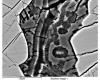
Initial Impression of the Micrograph: A preliminary viewing reveals elongated, somewhat parallel, and aggregated (bundled) structures. Their coloration, predominantly in shades of brown and orange, is suggestive of mineral staining, a common characteristic of fossilized materials. The elongated and parallel arrangement is a hallmark of many biological tissues designed for transport or structural support, particularly in plants. Wood (secondary xylem) is primarily composed of such axially elongated cells. The “bundled” appearance suggests a cohesive tissue rather than random mineral formations. This initial observation justifies a deeper investigation into a biological, and specifically woody, origin.
2. Detailed Visual Analysis of the Micrograph
Dominant Structural Features:
The micrograph is characterized by numerous elongated elements, appearing as strands or fibers. These elements are generally oriented in a parallel to sub-parallel fashion, creating a distinct “grain” or linear texture across the field of view. There is an apparent three-dimensionality to some structures, with some strands seemingly overlying others. This suggests a degree of preserved volume, which is consistent with permineralization processes. Permineralized fossils often retain the original three-dimensional shape of the material that was fossilized, rather than being flattened, because the infilling minerals provide internal support.1
Potential Cellular Characteristics:
Upon closer examination of the strands, individual units or compartments can be discerned, suggestive of cellular organization. These units appear elongated and are bounded by what seem to be preserved cell walls. The internal spaces (lumens) of these potential cells appear to be filled with a translucent to semi-translucent mineral. The quality of preservation is critical in identifying such features. Silicification, a common mode of wood fossilization, can preserve cellular detail to varying degrees. The best preservation is often found in specimens where cell interiors (lumina) remain empty, or, significantly, if the “cell lumina are filled with transparent quartz, it may be possible to examine internal pits, rays, and other anatomical details by transmitted light microscopy”.2 The micrograph appears to have been captured using transmitted light, and the somewhat transparent nature of the filled lumens aligns with this possibility of quartz infill. The outlines of these potential cells, interpreted as cell walls, are visibly darker or more defined than the infilling material, further delineating the cellular structure.
Coloration and Texture:
The predominant colors observed are various shades of orange, reddish-brown, and yellowish-brown. These colors are not uniform; there are variations and mottling within the structures. This coloration is highly significant. Petrified wood is colored according to the minerals that have been introduced into its cellular cavities, with the specific hues often depending on the presence of various metal oxides in the soil and groundwater during fossilization.3 Specifically, “Iron oxide ➡️ red,” and “Hematite ➡️ red and pink” are listed as common colorants.3 The observed coloration in the micrograph strongly suggests the presence of iron oxides, which are common accessory minerals in silicified wood, imparting these characteristic hues. The texture within the filled lumens appears somewhat crystalline or glassy, consistent with mineral infill such as silica in the form of quartz or chalcedony. Chalcedony is described as a cryptocrystalline variety of silica with a waxy luster.1 The remnants interpreted as cell walls appear more opaque or darker than the infilling mineral.
Surrounding Matrix:
There is no clearly discernible separate matrix material; the entire field of view seems to consist of these organized, mineralized structures. This suggests that the original tissue was relatively pure and that the fossilization process primarily involved the infiltration and alteration of the wood itself.
Absence of Obvious Non-Biological Artifacts:
The regularity and organization of the elongated structures argue against them being purely inorganic mineral growths (e.g., acicular crystals forming randomly), which typically lack the consistent orientation and cellular-like compartmentalization observed. The combination of organized structure and specific coloration points strongly to permineralized biological tissue, likely plant-derived. The view is almost certainly a longitudinal section of the original material, as the elongated appearance of the potential cells and their parallel arrangement is characteristic of how wood cells (tracheids, vessel elements, fibers) appear when sectioned along their length. A transverse (cross-sectional) view would show these cells as more circular or polygonal outlines.
3. Comparative Wood Anatomy: Correlating Micrograph Features with Known Wood Structures
Fundamental Wood Cell Types (Secondary Xylem):
To assess whether the structures in the micrograph are indeed fossilized wood cells, a comparison with the fundamental cell types found in wood (secondary xylem) is essential. Wood is primarily composed of:
Tracheary Elements: These are the principal water-conducting cells and also provide structural support. Mature xylem tissue comprises tracheary elements, fibers, and parenchyma cells. Tracheary elements are typically dead at maturity, arranged axially to form long pathways for sap transport, and possess thick, lignified secondary cell walls that resist collapse under tension.4
Tracheids: These are “relatively narrow conductive elements, connected to each other via holes in the secondary cell wall, called bordered pits.” Tracheids are a “major component of the wood in gymnosperms (conifers)” and also contribute to structural support in these plants, which generally lack specialized fiber cells.4 Softwoods (gymnosperm wood) are composed of 90 to 95 percent tracheids.5 Tracheids are typically elongated, spindle-shaped cells.
Vessel Elements: These are the “major conductive cell type in angiosperms, usually wider in diameter than tracheids and arranged axially, one above the other, to form long tubes called vessels”.4 When sectioned and viewed on the wood surface, vessels often appear as pores.5
Fibers: These are “long, mostly dead cells with lignified, thick secondary cell walls” and are the primary “supporting elements in angiosperm wood”.4 Most hardwood (angiosperm) tissue is composed of fibers.5
Parenchyma Cells: These cells remain living for many years within the wood and are involved in functions such as storage of carbohydrates and radial transport. They can be oriented axially (axial parenchyma) or radially (forming xylem rays).4 Of particular relevance to the user’s query, axial parenchyma cells “usually form strands of axially elongated cells”.4
Correlation with Micrograph Observations:
Elongated, Parallel Units: The dominant structures in the micrograph are highly consistent with the longitudinal appearance of axially oriented wood cells such as tracheids, fibers, or potentially narrow vessel elements. Their parallel arrangement directly reflects the typical grain of wood.
Cellular Dimensions (Qualitative): The visible cell-like units in the micrograph appear relatively narrow and fairly uniform in width. While precise measurement is not possible without a scale bar, there are no obviously very wide, pore-like structures that would definitively indicate the presence of large vessel elements typical of many angiosperm woods.
“Strands” and Axial Parenchyma: The user’s term “strands” could refer to the overall bundled appearance of the tracheary elements or fibers. Alternatively, and more specifically, if some of these “strands” were subtly distinct from the main conductive elements (e.g., composed of shorter cells or cells with different wall thickness or contents, though this level of differentiation is challenging to confirm from this single micrograph), they could represent fossilized axial parenchyma. As noted, axial parenchyma is described as forming “strands of axially elongated cells”.4 The observed structures strongly resemble longitudinally sectioned tracheids, which are characteristic of gymnosperm wood. The cells are elongated, relatively narrow, and appear fairly uniform in diameter. This morphology is highly consistent with tracheids.
Potential Absence of Angiosperm Features: The apparent lack of distinct, large-diameter vessel elements (which appear as pores in cross-section 5) might suggest the wood is not from a typical angiosperm (hardwood). However, it is important to note that some angiosperms possess very small vessels, or the plane of section in the micrograph might not intersect larger vessels if they were present. Nevertheless, the overall impression is one of greater homogeneity than is often seen in angiosperm woods.
Gymnosperm (Softwood) vs. Angiosperm (Hardwood) Considerations:
The structural differences between gymnosperm and angiosperm wood are significant. Gymnosperm wood is anatomically simpler, “primarily consists of tracheids (reaching up to 90% in volume) and only a small amount of xylem parenchyma cells, often limited to rays”.4 Softwoods, therefore, have a much simpler cellular structure, with 90 to 95 percent of the wood composed of tracheids; they lack fibers and vessel elements.5 In contrast, angiosperm wood is more complex, typically containing “vessel elements, tracheids, fibres, and abundant xylem parenchyma fractions”.4
The relatively uniform appearance of the elongated cells in the micrograph, if interpreted as predominantly one cell type (narrow tracheids), would be more characteristic of gymnosperm wood. Given that gymnosperm wood is overwhelmingly composed of tracheids 4, this observation leans the identification towards a gymnosperm. The absence of obvious large vessels further supports this interpretation, as vessels are a defining feature of most angiosperms. The term “strands” used by the user could directly refer to the visible bundles of these tracheid-like cells. While the possibility of these being axial parenchyma strands exists, distinguishing them with certainty from the dominant tracheid-like cells is difficult from this single longitudinal view without clear differentiation in cell morphology or contents.
The preservation of cellular outlines and the infilling of what appear to be cell lumens point directly to the process of permineralization. This mode of fossilization “is the infilling of natural pores in original organic material by minerals,” which “forms a three-dimensional internal mold of the original organic tissue, sometimes preserving details at the microscopic or cellular level”.1 Indeed, “The most informative kind of fossil is permineralization, in which the minerals fill cell cavities and conserve the original cell walls of the plant”.6 This description accurately reflects the features observed in the micrograph: structures interpreted as cell cavities (lumens) are filled with a secondary mineral, and the positions of the original cell walls are still evident as darker boundary lines.
Evidence for Silicification:
The glassy or crystalline appearance of the infilling mineral, combined with the characteristic orange, brown, and reddish iron oxide staining previously discussed 3, strongly suggests that the permineralizing agent was silica (SiO2). This process is known as silicification. “Silicification begins when silicic acid dissolved in groundwater precipitates on cell walls (Fig. 1). This amorphous silica is termed Opal-A. Eventually, Opal-A converts to Opal-CT, a microcrystalline mixture of cristobalite and tridymite that ultimately may become transformed to chalcedony, a microcrystalline variety of quartz”.2 The relative clarity and translucency of the infill in the micrograph suggest it might be a form of microcrystalline quartz or chalcedony, rather than opaque opal or heavily included material which might obscure cellular details more significantly.
Quality of Cellular Preservation:
The quality of cellular preservation in the micrograph appears to be good to very good. The boundaries interpreted as cell walls are relatively distinct, and the lumens are clearly infilled. There is no evidence of significant crushing or distortion of the cells, indicating that mineralization occurred prior to or prevented major compaction. The “degree of preservation of cellular architecture is variable” in fossil wood.2 While “The best preservation is commonly found in specimens where the cell interiors (lumina) remain empty” 2, excellent detail can also be preserved when lumens are filled with transparent minerals. In the micrograph, the lumina are filled, but the transparency of the infilling mineral allows the cellular structure to be observed, consistent with descriptions of quartz-filled lumina where “it may be possible to examine internal pits, rays, and other anatomical details by transmitted light microscopy”.2
Mineral Infill and Coloration:
The prominent orange, brown, and reddish colors are indicative of iron compounds (such as goethite or hematite) that were likely precipitated along with or subsequent to the silicification process.3 These iron minerals often stain the silica or become incorporated as inclusions within it, contributing significantly to the final appearance of the fossilized wood. The fossilization process is therefore clearly permineralization by silica, with associated iron mineralization responsible for the observed coloration. The clarity of the infill suggests a later stage of silicification, potentially involving the formation of microcrystalline quartz, as opposed to early-stage amorphous opal, which can sometimes obscure details if it is not well-ordered or if crystal sizes are large and poorly formed.2
5. Contextual Clues: “fossil_lin” and Potential Specimen Origin
Investigating “fossil_lin”:
The username “fossil_lin” strongly suggests an individual involved in fossil collection or paleontological research. Searches for this name in the context of fossil studies reveal connections to paleontological work, particularly in Taiwan. Multiple publications link the surname “Lin” to paleontological research in this region. For instance, Chien-Hsiang Lin is listed as an author on several papers concerning Taiwanese fossils, including material from the Liuchungchi Formation.7 Another search result links “fossil_lin” to “Lin De Kai” and a paper on Keteleeria fossil wood from South China.11 While the exact identity of “fossil_lin” cannot be definitively established from the available information alone, the association with Taiwanese paleontology, particularly through Chien-Hsiang Lin’s work, appears to be a strong possibility.
Fossil Wood from Taiwan:
Taiwan has documented occurrences of fossil wood. Notably, “fragments of carbonized woods are also known” from the Niubu area, Chiayi, southwestern Taiwan, within the Early Pleistocene Liuchungchi Formation.7 This formation is a key research area for Chien-Hsiang Lin. The term “carbonized woods” in field reports might be a general descriptor for any dark-colored fossil wood encountered before detailed laboratory analysis. True carbonization results in a coal-like material rich in carbon. However, wood can undergo partial carbonization and then be subsequently permineralized, often by silicification. The micrograph under examination clearly shows features consistent with silicification (mineral infill, translucency, iron staining) rather than primary carbonization. It is plausible that the specimen underwent an initial phase of partial carbonization, which may have darkened the cell walls, followed by silicification that preserved the structure. Alternatively, “carbonized” might be used loosely in some contexts for any dark, organic-rich fossil wood. The visual evidence in the micrograph points to silicification as the dominant and final mode of preservation visible.
Further evidence of fossil wood research in the region includes a paper by Lo and Hu describing a new fossil wood species, Podocarpoxylon nageioides (a conifer type), from Miocene strata in Northern Taiwan.13 Additionally, the aforementioned study potentially linked to “Lin De Kai” describes Keteleeria sp. fossil wood, a conifer in the Pinaceae family, from the Late Pleistocene of South China, a region geographically proximal to Taiwan.11
Implications of Potential Taiwanese Origin:
If the specimen depicted in the micrograph is indeed from Taiwan, particularly from formations like the Liuchungchi Formation, the presence of fossil wood is well-documented. The types of wood reported from the broader region (e.g., conifer woods such as Podocarpoxylon or Keteleeria-like woods) would be consistent with the gymnosperm-like features tentatively observed in the micrograph (e.g., relatively uniform tracheids, lack of obvious large vessels). The geological settings described for these formations, such as marine sedimentary environments that received terrestrial input like wood fragments 7, are conducive to the fossilization of wood through processes like silicification, which typically involves silica-rich groundwater.2 The strong possibility that “fossil_lin” is a Taiwanese paleontologist and that the image is of a Taiwanese fossil wood specimen significantly contextualizes the find. Researchers often share images of their study material, and if “fossil_lin” is indeed Chien-Hsiang Lin, the micrograph is likely from a site he has worked on. This does not alter the primary visual interpretation but adds substantial weight to the identification as fossil wood, as such material is expected from that geological and geographical context.
6. Synthesis and Conclusion
Summary of Evidence:
The micrograph under investigation displays numerous elongated, parallel, and compartmentalized structures. These features are highly consistent with the appearance of wood cells when viewed in a longitudinal section. The structures are infilled with a translucent mineral, and they exhibit coloration in shades of brown, orange, and red, which is characteristic of iron oxides commonly associated with silicified fossil wood. The overall morphology, particularly the relatively uniform diameter and elongated nature of the cell-like units, strongly aligns with that of tracheids, the predominant cell type in gymnosperm (softwood) wood.
Assessment of User Query:
Based on the comprehensive visual analysis of the micrograph, and through comparison with established knowledge of wood anatomy and common fossilization processes, it is highly probable that the micrograph indeed shows strands of fossilized wood cells. The term “strands” aptly describes the bundled appearance of these longitudinally oriented cellular elements.
Probable Nature of the Wood:
The uniformity of the narrow, elongated cells and the apparent absence of large, distinct vessel elements (which are characteristic of most angiosperm woods) suggest that the wood is likely derived from a gymnosperm (conifer). In this interpretation, the “strands” are primarily composed of bundles of fossilized tracheids.
Fossilization Process:
The mode of preservation is clearly permineralization, specifically silicification, where silica (SiO2) has infilled the cell lumens and possibly replaced original cell wall material to some extent. The prominent coloration is attributed to significant iron mineralization occurring concomitantly with or subsequent to the silicification.
Contextual Support:
The potential origin of the image from “fossil_lin,” who may be a Taiwanese paleontologist, and the known occurrences of fossil wood (including conifer wood) in Taiwanese geological formations, such as the Liuchungchi Formation 7, lend further contextual support to this identification.
Table 1: Comparison of Micrograph Features with Diagnostic Characteristics of Fossilized Wood
Axially oriented cells like tracheids, fibers, or narrow vessel elements in longitudinal section; cell lumens and walls
Consistent with wood cellular structure; suggests preservation of original biological organization.
Translucent mineral infill in cell lumens
Cell lumens (internal cavities of cells)
Indicates permineralization, where minerals have filled empty spaces within the cells.
Brownish-orange-reddish coloration
N/A (coloration is a feature of fossilization, not original wood color in this context)
Strongly suggests iron oxide mineralization (e.g., hematite, goethite), common in silicified wood.3
Relatively uniform, narrow cell diameter
Tracheids (typical of gymnosperms) or small-diameter fibers/vessel elements (in some angiosperms)
Consistent with tracheids, which are the dominant cell type in gymnosperm wood 4; leans towards a gymnosperm origin.
Apparent absence of wide, distinct vessel elements
Absence of large pores (vessels)
Supports gymnosperm wood identification, as most gymnosperms lack vessels 5; some angiosperms also have small/absent vessels.
The convergence of multiple lines of evidence—morphological characteristics consistent with wood cells, coloration indicative of specific mineralization processes common in fossil wood, anatomical features aligning with gymnosperm wood structure, and plausible contextual information regarding the image’s source—provides high confidence in the identification of the features in the micrograph as strands of fossilized wood cells, likely of coniferous origin.
Limitations:
It is important to acknowledge that this assessment is based solely on the analysis of a single two-dimensional micrograph. A definitive identification to a specific wood genus or species, or a more detailed anatomical characterization, would necessitate the examination of physical specimens. This would ideally include the preparation and study of thin sections in multiple orientations (transverse, radial, and tangential) to observe critical diagnostic features such as the type and arrangement of pitting on cell walls, the detailed structure of xylem rays, and the distribution of axial parenchyma, none of which are clearly discernible in the provided longitudinal view.
7. Further Considerations
For the Image Owner/User with Access to the Specimen:
Should a physical sample corresponding to this micrograph be available, further microscopic examination could yield significantly more detailed information and allow for a more precise identification. Recommended procedures would include:
Preparation of Thin Sections: Creating polished thin sections of the fossil wood in three standard anatomical planes—transverse (cross-section), radial longitudinal, and tangential longitudinal—is crucial. Different anatomical features are best observed in specific planes. For instance, transverse sections are vital for observing the overall arrangement of cells, the presence and distribution of vessels (if any), growth ring characteristics, and axial parenchyma patterns.14 Radial sections are optimal for examining details of pitting on the radial walls of tracheids and the structure of cross-fields (the interface between axial tracheids and ray cells). Tangential sections provide information on the height and width of xylem rays and the arrangement of cells within them.4 The study of such sections is standard practice in paleoxylology (the study of fossil wood).13
Polarized Light Microscopy: Observing the thin sections under a polarizing microscope can enhance the visibility of crystalline structures within the permineralizing silica and can help delineate fine details of cell wall layering or alteration.15
Scanning Electron Microscopy (SEM): If higher magnification and resolution are needed to examine very fine details, such as pit membranes or minute surface textures of cell walls, SEM could be employed. However, as noted in studies of silicified wood, SEM is most effective for revealing surface topography, and its utility might be reduced if cell lumens are completely filled with highly transparent quartz, which minimizes topographic contrast.2 Transmitted light microscopy often provides superior views of internal structures in such cases.
General Note on Micrograph Interpretation:
While the provided micrograph offers compelling evidence for the presence of fossilized wood cells, it is a standard principle in paleobotany that robust identifications, particularly to lower taxonomic levels (genus or species), rely on the comprehensive analysis of a suite of anatomical characters observed from multiple perspectives (i.e., different section planes). The current analysis confidently addresses the user’s specific query regarding the presence of fossilized wood cells. However, to advance the identification further, for example, to determine the family or genus of the conifer, would necessitate the more comprehensive paleobotanical preparation and analytical techniques outlined above.
Before It’s News® is a community of individuals who report on what’s going on around them, from all around the world.
Anyone can join.
Anyone can contribute.
Anyone can become informed about their world.
"United We Stand" Click Here To Create Your Personal Citizen Journalist
Account Today, Be Sure To Invite Your Friends.
Before It’s News® is a community of individuals who report on what’s going on around them, from all around the world.
Anyone can join.
Anyone can contribute.
Anyone can become informed about their world.
"United We Stand" Click Here To Create Your Personal Citizen Journalist Account Today, Be Sure To Invite Your Friends.
Mushrooms are having a moment. One fabulous fungus in particular, lion’s mane, may help improve memory, depression and anxiety symptoms. They are also an excellent source of nutrients that show promise as a therapy for dementia, and other neurodegenerative diseases. If you’re living with anxiety or depression, you may be curious about all the therapy options out there — including the natural ones.Our Lion’s Mane WHOLE MIND Nootropic Blend has been formulated to utilize the potency of Lion’s mane but also include the benefits of four other Highly Beneficial Mushrooms. Synergistically, they work together to Build your health through improving cognitive function and immunity regardless of your age. Our Nootropic not only improves your Cognitive Function and Activates your Immune System, but it benefits growth of Essential Gut Flora, further enhancing your Vitality.
Our Formula includes:
Lion’s Mane Mushrooms which Increase Brain Power through nerve growth, lessen anxiety, reduce depression, and improve concentration. Its an excellent adaptogen, promotes sleep and improves immunity.
Shiitake Mushrooms which Fight cancer cells and infectious disease, boost the immune system, promotes brain function, and serves as a source of B vitamins.
Maitake Mushrooms which regulate blood sugar levels of diabetics, reduce hypertension and boosts the immune system.
Reishi Mushrooms which Fight inflammation, liver disease, fatigue, tumor growth and cancer. They Improve skin disorders and soothes digestive problems, stomach ulcers and leaky gut syndrome.
Chaga Mushrooms which have anti-aging effects, boost immune function, improve stamina and athletic performance, even act as a natural aphrodisiac, fighting diabetes and improving liver function.
Try Our Lion’s Mane WHOLE MIND Nootropic Blend 60 Capsules Today. Be 100% Satisfied or Receive a Full Money Back Guarantee. Order Yours Today by Following This Link.